Artificial intelligence in medicine: Getting smarter one patient at a time

What if doctors had more time to spend addressing their patients' concerns? That's the thrust behind the push for integrating artificial intelligence (AI) into medicine. By using complex algorithms to detect patterns in large datasets—like lab test results, current medications, and symptoms, to name a few—AI might actually make medicine more personable—not less.
"By the time patients come in, we would already know what they've been experiencing," says Yale Medicine cardiologist and data researcher Harlan Krumholz, MD. "Especially for patients with chronic conditions, we could detect their need for medical attention before they do."
The definition of AI varies among industries and even from one dictionary to another. But broadly speaking, in the realm of medicine, AI refers to the use of computer systems to create algorithms based on patterns in raw data to find connections (such as between a genetic mutation and a medical condition—or clusters of symptoms to a particular disease) that would be very hard, if not impossible, for a person to identify.
To illustrate what an AI-assisted future in medicine will look like, Dr. Krumholz gives a hypothetical example of a patient at risk of heart failure, a condition where a weakened heart muscle struggles to pump enough oxygenated blood throughout the body.
To start, the patient would start the day by stepping on an internet-connected scale that would monitor changes in weight for possible signs of fluid retention—a hallmark of heart failure. He or she would strap on a smart watch or other sensor to track steps and activity level, and would use a phone app to log specific symptoms, such as shortness of breath. All of this data would stream directly to the electronic health record (EHR), which would "take all of that information and categorize the patient's risk, rather than wait for the patient to come to us," Dr. Krumholz says. The doctor then could alert the patient that he or she is approaching danger and take steps to avert it, allowing medical care to be given proactively rather than reactively.
This kind of AI-assisted interaction in medicine has been years in the making, Dr. Krumholz explains. Since 1995, he has directed the Center for Outcomes Research and Evaluation (CORE), whose research has helped improve patient care by gathering, measuring, and analyzing all kinds of data, from billing records to traditional medical records and now EHRs.
Before patients' records were digitized and able to be analyzed by computers via algorithms and machine learning, Dr. Krumholz and his team worked to extract insights from the paper records. This expensive, labor-intensive work took years to complete. The research—a result of a collaboration with federal agencies, medical professional organizations such as the American College of Cardiology and the American Heart Association, as well as clinicians, hospitals, and others—led to dramatic improvements in care. In one notable effort, which spanned years, the research and its dissemination led to drastically reducing the time it takes for heart attack patients to receive life-saving treatment that clears their blood clots.
Today, such projects could be accomplished requiring only a fraction of the time and resources. As Dr. Krumholz notes, "In this digital era, the prospect of real-time research producing real-time actionable information and timely improvements in care is just around the corner."
AI and precision medicine
The Food and Drug Administration (FDA) now evaluates AI tools in much the same way they review drugs and medical devices for efficacy and safety. For example, one approved AI system analyzes CT scans of patients presenting with neurological symptoms, texting doctors when the results suggest stroke. This helps them deliver effective treatment faster, which in turn helps prevent brain damage from strokes.
"Machine learning and AI help us put together highly complex, high-dimensional data in ways that we just couldn't do before with our more traditional analytics," Dr. Krumholz says.
Here are some of the ways AI is currently being used by Yale Medicine doctors to bring faster, more effective treatments to patients.
Machine learning and prostate cancer
Even though prostate cancer is the second most common kind of cancer in the U.S., it remains challenging to diagnose. Typically doctors must repeatedly insert biopsy needles into the prostate, gathering several samples from the area a mass is thought to be in the hope they will obtain a cluster of the cancerous cells. Because the prostate is a soft organ that can rotate and move within the pelvis, "It's a very difficult thing to biopsy," explains Yale Medicine urologist Preston C. Sprenkle, MD.
Using AI-assisted machine learning, Dr. Sprenkle combines ultrasound and MRI scans from a patient into one image that shows more precisely where the suspected tumor is located. "Both the MRI and the ultrasound show very different visualizations of the anatomy, and the challenge is trying to map those two images together," says John Onofrey, Ph.D., assistant professor of radiology and biomedical imaging and of urology, who worked with Dr. Sprenkle to set up the AI tool.
The tool, called the MRI/TRUS Fusion for Prostate Biopsy, gathers image data and feeds that information into an algorithm that then creates a 3-D image of the prostate. Doctors can then explore these images from different angles on a computer screen in order to precisely identify lesions to target for a biopsy.
"We've learned from hundreds of cases that have come prior to this, and with some manual training from Dr. Sprenkle, how the prostate can deform [or change shape] during this biopsy procedure," Onofrey says. By first mapping images with AI software, doctors are able to do more precise—and therefore fewer—insertions of needles to retrieve biopsy samples, which, as one can imagine, dramatically improves the experience of the patient undergoing the procedure.
Artificial intelligence and liver cancer
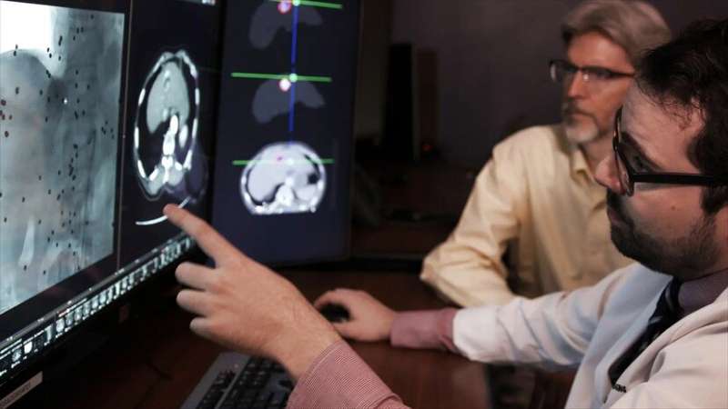
Liver cancer is complex, so doctors need to consider information from many different sources to determine how best to treat a particular patient. For example, to see the cancer, they rely on CT and MRI images. They must measure the tumor's size and try to understand how quickly it is growing. They try to identify particular genes found within the tumor and also weigh the patient's medical and family history to help guide treatment plans.
Working to find ways to improve accuracy by incorporating additional and disparate data points, Yale Medicine liver cancer specialists approached a Yale team of biomedical engineers and computer scientists to explore the creation of an algorithm that could help them recognize patterns in the data.
"We looked to the clinicians to give us the appropriate clinical questions," explains Lawrence Staib, Ph.D., a radiology and biomedical imaging researcher, who specializes in using machine learning to analyze medical images. "Then, it's a narrative process of testing algorithms and evaluating how well they're performing."
Both sides focused on getting better at recognizing patterns in images.
"In liver cancer, imaging plays a very, very important role," says Yale Medicine researcher and interventional radiologist Julius Chapiro, MD. "We need to get better at extracting the imaging information in a quantitative way."
The AI-produced algorithms are proving helpful in bridging a gap between complex data and clinical decision-making. Though there is still room for improvement, the team is already seeing advantages to this approach.
"Humans are imperfect," says Jeffrey Pollack, MD, another interventional radiologist involved in the project. "And machines aren't going to be perfect, either. But maybe putting the two together will achieve a greater level of perfection."
3-D planning for facial plastic surgery
Machine learning techniques can add another level of accuracy to computer-assisted 3-D design models, which help plastic surgeons prepare for complex facial reconstruction surgery.
"There's so many functional considerations that go along with the face," says Derek Steinbacher, DMD, MD, a Yale Medicine plastic and reconstructive surgeon. "Most of the time, it's form-function," he says, referring to the need for structures, like the delicate bones of the human face, to have balanced proportions in order to function well.
By collaborating with researchers nationally and globally, Yale Medicine surgeons created a machine learning model based on images of about 4,000 people with normal facial structures. Working within the specialized field of morphometrics, which relies on measuring and testing factors that affect the shape or form of living organisms using quantitative analysis, the team created 3-D models of faces.
Computer programs then compare large amounts of normal facial models with ones made from post-operative scans, providing insight surgeons can use to improve surgery results.
"We could use a model [to understand] what the bone relationship will be and how we need to move the bone to achieve a facial result," says Dr. Steinbacher, who directs Yale Medicine's craniofacial surgery program and is chief of oral maxillofacial surgery and dentistry.
To build a model, physicians compile MRI and other imaging scans from a patient's medical record. "We can then render them digitally and basically perform the surgery in a digital space," Dr. Steinbacher explains. Once the 3-D model is accurate, surgeons might print a model of a patient's facial bones and use this in the operating room to guide real-time surgery.
"I think incorporating this model into our planning process will help us get reproducible, high-fidelity, and accurate results," Dr. Steinbacher says.
More information: The Center for Outcomes Research and Evaluation: medicine.yale.edu/core/


