This article has been reviewed according to Science X's editorial process and policies. Editors have highlighted the following attributes while ensuring the content's credibility:
fact-checked
trusted source
proofread
Virtual dissection fleshes out instruction in animal science anatomy lab

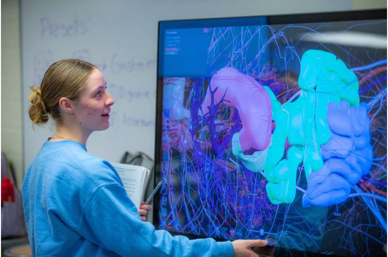
In a recent class session devoted to reviewing the components of a monogastric digestive system, Alexandra Else-Keller reminded an animal science student how to position her fingers as they examined how the colon, stomach, and gallbladder nestle together.
"One finger rotates it. Two fingers move it," she said, describing how to manipulate a 3D digital rendering of a bowel system to see it from an angle with a better view of the gallbladder.
A virtual dissection table is a relatively common teaching tool in medical and nursing schools, but it's a rare opportunity for animal science students. The 180 students per semester who take Domestic Animal Physiology, the foundational anatomy lab in Iowa State University's animal science department, use the technology to supplement their hands-on learning via more traditional methods such as examining fresh tissues, preserved specimens, and plastic models.
"It's been super helpful. You can say to students, "OK, show me where this is,'" said Else-Keller, a doctoral student who is one of the course's head teaching assistants. "You want to give students with different learning styles multiple ways to learn because not everyone learns the same way."
Including an interactive digital component in the anatomy lab curriculum is a point of pride for the department, said Karl Kerns, an assistant professor of animal science who oversees the course.
"We're the first and only animal science department and pre-veterinary program in the nation, to our knowledge, using a virtual anatomy table for classroom instruction," Kerns said.
Virtual dissection has been used in the course for two years, accessed either through a large main central touchscreen or by tablets that connect to monitors throughout the Kildee Hall classroom lab. It doesn't replace other methods of studying animal physiology, Kerns said. The benefit is getting a fuller perspective on where organs are in a body and how they work.
"You can literally see how the blood flows through the heart, with the valves opening and closing. You can see an ECG chart and side-by-side which compartment of the heart is beating in real time to create the electrogram. You can interact with it," he said.
Sydney Plumb, a sophomore in animal science taking the anatomy lab this semester, said virtual dissection helps reinforce what she learns in class and better understand the positioning of internal body systems.
"A picture can only tell you so much," she said.
There are some animal models loaded into the system, and more may be added in the future, but for now, students mostly study a fully annotated human body. That's just as useful earlier in the semester when students learn anatomy basics such as the cardiovascular system and central nervous system, before moving on to animal-specific lessons later in the semester, Kerns said.
"A heart is a heart, whether it's a pig or a cow or a human," he said.


