New AI solutions help scientists track objects in images; will aid discoveries in life sciences
New AI solutions help scientists track objects in images; will aid discoveries in life sciences
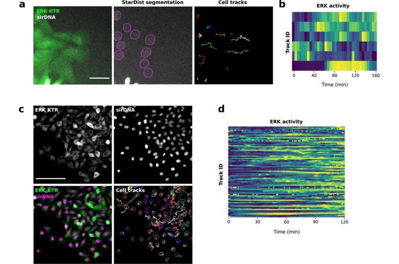
U2OS (a, b.) and MDA-MB-231 cells (c. and d.) stably expressing an ERK activity reporter (ERK-KTR-Clover) and labeled using SiR-DNA were recorded live using a widefield fluorescence microscope. U2OS cells were recorded live over 3 hours (1 image every 5 minutes) and MDA-MB-231 cells were recorded live over 2 hours (1 image every minute). Cell nuclei were automatically tracked over time by using StarDist in TrackMate. A custom StarDist model was trained to detect the U2OS nuclei using the ZeroCostDL4Mic platform. The “Versatile fluorescent nuclei” StarDist model was used to track the MDA-MB-231 cell nuclei. For each tracked cell, the average intensity of the ERK reporter was measured in their nucleus over time (directly in TrackMate). Changes in ERK activity are displayed as heatmaps (blue high, yellow low). Heatmaps were generated using PlotTwist. Scale bar = 250 µm. Credit: Dmitry Ershov et al, Nature Methods (2022). DOI: 10.1038/s41592-022-01507-1

Object tracking⁠—following objects over time⁠—is an essential image analysis technique used to quantify dynamic processes in biosciences. A new application called TrackMate v7 enables scientists to track objects in images easily. TrackMate is a free, open-source tool available as part of the Fiji image analysis platform.

"TrackMate allows scientists to tackle complex tracking problems more efficiently, accelerating discoveries in across fields," says Guillaume Jacquemet, Academy Research Fellow at Åbo Akademi University and one of the researchers involved in TrackMate development.

In life sciences, tracking is used, for instance, to follow the movement of molecules, subcellular organelles, bacteria, cells, and whole animals. However, due to the sheer diversity of images used in research, no single application can address every tracking challenge.
New AI solutions help scientists track objects in images; will aid discoveries in life sciences
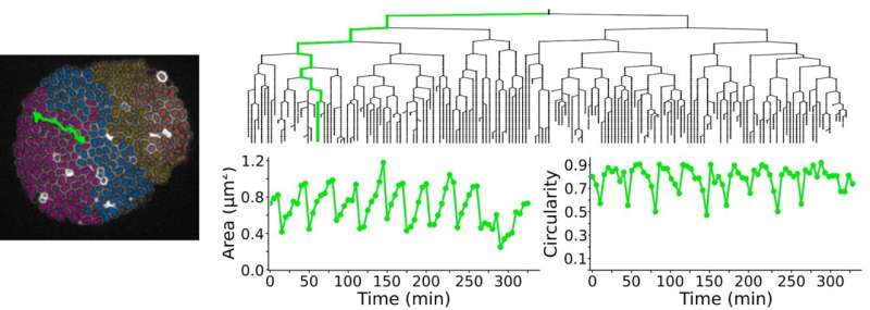
Bacteria growth (Neisseria meningitidis) was followed over time using TrackMate v7. A track and lineage of a single bacterium are highlighted in green, and changes in bacteria shape (area and circularity) over the tracking period were plotted. Bacteria division can be observed through the dramatic changes in the area and circularity. Credit: Nature Methods (2022). DOI: 10.1038/s41592-022-01507-1

TrackMate v7 offers automated and semi-automated tracking algorithms and advanced visualization and analysis tools. To analyze a wide variety of images, the application relies on artificial intelligence solutions and other advanced segmentation algorithms to detect objects from images.

"This new feature widely increases the breadth of TrackMate applications and capabilities. For instance, we show that TrackMate v7 can be used to follow moving cancer cells, immune cells, or stem cells. It can also be used to follow bacteria growth," says Jacquemet.

"We are currently using the software in the Jacquemet laboratory to study the mechanisms enabling cancer metastasis. We can now produce better and more informative data much faster than ever before," he adds.

The development of TrackMate v7 was coordinated by the Jacquemet (Åbo Akademi University, Turku, Finland) and Tinevez laboratories (Pasteur Institute, Paris, France).

Their research is published in Nature Methods.

More information: Dmitry Ershov et al, TrackMate 7: integrating state-of-the-art segmentation algorithms into tracking pipelines, Nature Methods (2022). DOI: 10.1038/s41592-022-01507-1

Journal information: Nature Methods
Provided by Abo Akademi University
Citation: New AI solutions help scientists track objects in images; will aid discoveries in life sciences (2022, June 2) retrieved 19 April 2024 from https://techxplore.com/news/2022-06-ai-solutions-scientists-track-images.html
This document is subject to copyright. Apart from any fair dealing for the purpose of private study or research, no part may be reproduced without the written permission. The content is provided for information purposes only.

Explore further

An easy-to-use platform is a gateway to AI in microscopy

63 shares

Feedback to editors