This article has been reviewed according to Science X's editorial process and policies. Editors have highlighted the following attributes while ensuring the content's credibility:
fact-checked
trusted source
proofread
A peek inside art objects: New algorithm makes CT scan more accessible

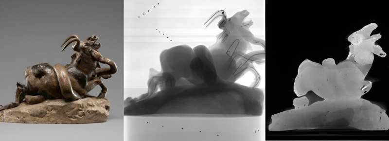
An X-ray scanner, some small metal balls, and a newly developed algorithm. That is all you need to make a 3D model that enables you to look inside art objects without dismantling them. Thanks to the research of Francien Bossema (Centrum Wiskunde & Informatica and Leiden Institute of Advanced Computer Science), museums can now use existing X-ray equipment as CT scanners, without having to buy such a costly and complicated device.
What is on the inside of an art object? To answer that question, art experts can use an X-ray machine. Some museums own one for inspecting their objects. They use the machine to see whether an object has woodworm, for example, and to what extent. But such X-rays have drawbacks.
You see everything on top of each other with no depth, so you can never really make a cross-section of the object. A CT scanner can do that but not many museums can afford one. Bossema and her supervisor Joost Batenburg wondered: can we make better use of what we already have?
X-ray machine becomes aspiring CT scanner
A CT scanner is actually an X-ray scanner that captures the object from all angles. So you take hundreds or thousands of X-rays in a row. You then use a reconstruction algorithm to use those photos to create a 3D model of the object, which you can digitally slice in different directions.
With a professional CT scanner, as in a hospital, the knowledge of the exact position of all parts is automated. Bossema has now developed an algorithm to gather that knowledge after the scan has been made. Thus, a simple X-ray scanner becomes an aspiring CT scanner.
Metal balls as placeholders
We've heard about the X-ray scanner and the algorithm, but what about those small metal balls? Bossema said, "To make a CT scan, you need to be able to move the X-ray machine around the object. When you do that, you have to know exactly where everything was during the scan. Where is the source in relation to the turntable? How many degrees are we rotated between two X-rays? Where is the detector located? All these places you need to know very precisely. That's why we put small metal balls next to the object."
These balls have a very high density and become thick black dots on the X-ray photo. "We look for black dots on those X-rays, which naturally move when you turn the object. With these reference points, you can calculate how much the object has been rotated. If you know that for all the photos, you can construct a 3D image of the object," she added.
Building bridges between the beta and art world
Bossema tested the algorithm at four different locations, including three museums. At the Rijksmuseum in Amsterdam and the British Museum in London, she did the measurements herself. At the J. Paul Getty Museum in Los Angeles, she provided instructions only via e-mail and Zoom. With this, Bossema concludes that the method could be generally applicable.
She said, "If you know the Python programming language, you can basically use my software. But for art experts, it might be a bridge too far." A more accessible user interface could help, but that is beyond Bossema's research. She hopes someone will have the time and space to take the project further.
Building bridges between science and art research really attracts Bossema. "My research also really has a practical application. I have not only written my own articles about the algorithm and the technique behind it, but I also co-wrote articles by colleagues, because I have collaborated with my technique on projects by other researchers at the museum. I really like that, that my research in turn also facilitates the work of my colleagues," she explained.
For now, Bossema is not ready to leave the museum world. This summer, she will spend ten weeks working with CT scans at the Getty in Los Angeles, and she is also a postdoc fellow at the Rijksmuseum Amsterdam.
Besides mathematics, Bossema studied science communication. This helped her a lot during her Ph.D. research, she says. "This project involves a lot of communication because I work with people at the museum who have a very different background from mine. They often don't know what an algorithm is, or what a CT scanner can do for their work. I find it very much fun and important to understand what they need. Not everyone in mathematics finds this communication aspect interesting. So that does make me unique as a researcher."


